

Babies born to mothers who suffer from anaemia during pregnancy have total brain volumes approximately 4% smaller than those born to non-anaemic mothers, a study has found.
The research, led by King’s College London and published in the journal Brain Communications on 9 September 2026, highlights the critical role iron plays in foetal neurodevelopment. The study utilised novel ultra-low-field MRI technology to examine infants, making it easier to conduct detailed brain imaging in a wider range of settings.
The scale of anaemia in the UK
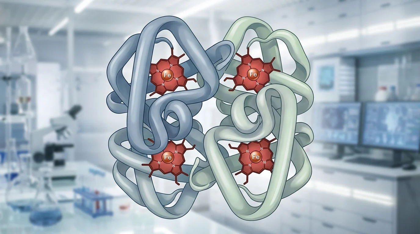
Iron deficiency anaemia is a significant public health issue in the UK, affecting approximately 30% of pregnant women. The condition occurs when the body lacks sufficient iron to produce haemoglobin, the protein in red blood cells responsible for carrying oxygen to the body’s tissues and the developing baby.
The study found that anaemia during pregnancy could lead to measurable differences in brain structure.
NHS screening and thresholds
In the UK, the NHS provides routine screening for anaemia to ensure pregnant women receive appropriate support and supplementation if required. Standard tests are typically carried out at the initial “booking” appointment (usually between 8 and 12 weeks) and again at 28 weeks of pregnancy.
Clinical guidelines define anaemia in pregnancy as a haemoglobin concentration below 110 g/L during the first trimester. In the second and third trimesters, this threshold is slightly lower, at 105 g/L. If levels fall below these marks, iron supplements are usually recommended alongside dietary changes.
Health officials advise that iron-rich foods can help maintain healthy levels. These include:
- Red meat
- Beans and lentils
- Dark green leafy vegetables, such as spinach and kale
- Fortified breakfast cereals
To improve iron absorption, the NHS suggests consuming vitamin C-rich foods or drinks, such as orange juice, alongside meals. Conversely, tea and coffee can hinder the body’s ability to absorb iron and are best avoided during meal times.

